
The major function of the reproductive system is to make sure that the human species survives. It is not necessary for every human being to produce children, but in order to continue our human species, at least some people have to have children. The four main functions of the human reproductive system are: To produce egg and sperm cells, to transport and sustain these cells, to nurture the developing offspring, and to produce hormones.
All of these functions are divided by their primary and secondary, or accessory, reproductive organs.
In the human reproductive system, the major organs include the external genitalia and many internal organs including gamete producing gonads. The external genitalia are the penis and vulva and the gamete producing gonads are the testicles and ovaries. These organs produce the egg and sperm cells (gametes) and produce hormones. These hormones are needed in the maturing of the human reproductive system, the development of sexual characteristics, and the importance in regulating the normal physiology of the human reproductive system. In the males, the major organ of the reproductive system is the penis, and in the females it is the vagina.
Female System
Female External Reproductive Organs
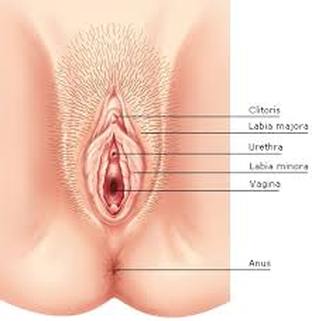
A female's external part of the reproductive organs is called the vulva, meaning “covering.” The vulva is located between the legs and covers the opening to the vagina and other reproductive organs located inside the body.
A female's external part of the reproductive organs is called the vulva, meaning “covering.” The vulva is located between the legs and covers the opening to the vagina and other reproductive organs located inside the body.
Female Internal Reproductive Organs
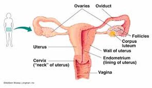
The female has four internal reproductive organs: the vagina; uterus; fallopian tubes; and, the ovaries.
Vagina
The vagina is a muscular, hollow tube that extends from the vaginal opening to the uterus. The vagina is about 3 to 5 inches long in a grown woman. The vagina has muscular walls and can expand and contract to become wider or narrower to accommodate the width of a baby. The vagina's muscular walls are lined with mucous membranes, which keep it protected and moist. The vagina has several functions: for sexual intercourse, as the pathway that a baby takes out of a woman's body during childbirth, and as the route for the menstrual blood (the period) to leave the body from the uterus. The vagina is where semen from the man is deposited during sexual intercourse. The vagina connects with the uterus at the cervix. The cervix has strong, thick walls and a very small opening, no wider than a straw. During childbirth, the cervix can expand up to 50 times its normal width to allow a baby to pass.
Uterus
The vagina connects with the uterus, or womb, at the cervix. The uterus is shaped like an upside-down pear, with a thick lining and muscular walls, and holds the developing baby during the nine months after conception. The uterus contains some of the strongest muscles in the female body. These muscles are able to expand to allow the uterus to grow 10 to 20 times its normal size during pregnancy and contract to push the baby out during labor. When a woman isn't pregnant, the uterus is only about 3 inches long and 2 inches wide.Each month the uterus goes through a cyclical change, first building up its endometrium or inner lining to receive a fertilized egg, then, if conception does not occur, shedding the unused tissue through the vagina in the monthly process called menstruation.
Fallopian Tubes
The female has two fallopian tubes, or oviducts, that attach to the upper corners of the uterus. The fallopian tubes are about 4 inches long and about the width of a piece of spaghetti. In each tube is a sewing needle size passageway. At the other end of each fallopian tube is a fringed area that looks like a funnel and wraps around the ovary. The fallopian tube does not completely attach to the ovary. When an egg pops out of an ovary, it enters the fallopian tube and tiny hairs in the tube's lining help push it down the narrow passageway toward the uterus. After intercourse, sperm in the vagina passes through the cervix, the uterus, and then to the fallopian tubes. If a sperm encounters an ovum (egg), conception occurs.
Ovaries
The female has two oval-shaped organs, called ovaries, that lie to the upper right and left of the uterus. The ovaries produce, store, and release eggs into the fallopian tubes during the process called ovulation. Each ovary measures about 1 1/2 to 2 inches in a grown woman. The ovaries are part of the endocrine system and produce estrogen and progesterone, female sex hormones. The ovaries are among the first organs to be formed as a female baby develops. By 20 weeks, the ovaries have about 6 to 7 million potential egg cells. From that point on however, the number begins to decrease rapidly. A newborn infant has between 1 million to 2 million egg cells. By puberty, the number of egg cells remaining is about 300,000. For every egg that matures and undergoes ovulation, roughly a thousand will fail. During the course of an average reproductive lifespan, roughly 300 mature eggs are produced for potential conception.
Male System
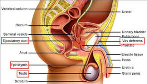
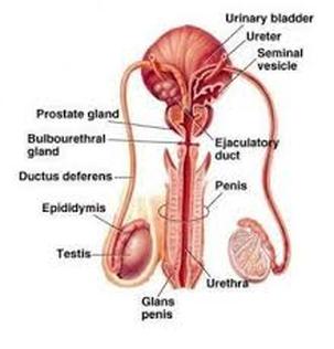
The male reproductive system is a complex system. Unlike the female system, the male reproductive organs, also known as genitales are located both inside and outside the pelvis. The male body parts include the following:
When a male who has reached sexual maturity, his testicles produce millions of tiny sperm cells. The testicles are oval shaped and are 2 inches long and 1 inch in diameter. The testicles are part of the endocrine system because they produce hormones such as testosterone. This is a major part of going through puberty and makes the male body go through some significant changes like body and facial hair, deeper voices, and larger muscles. The testicles are located in a sack-like body part called the scrotum. This bag of skin is used to regulate the temperature of the testicles and needs to be made colder than the rest of the body so sperm can be produced. It changes size to keep itself at the right temperature. Testosterone is the male sex hormone. The accessory glands, including the seminal vesicles and the prostate gland, provide fluids that lubricate the duct system and nourish the sperm. The prostate gland which produces some of the parts of semen surrounds the ejaculatory glands at the base of the urethra just below the bladder. The penis is actually made up of two parts: the shafts and the glands is the tip (also called the head). At the ends of the glands is a small slit or opening which is where semen and urine exit the body. The inside of the penis is made of spongy like material that can expand and contract.
All baby boys are born with a piece of skin at the tip called a foreskin located at the end of the penis covering the glands. Some males are circumcised which means the doctor or clergy member removes the baby's foreskin. This is usually done within the first few days after the baby has been born. Although circumcision is not medically necessary, parents who choose to have their children circumcised often do so based on religious beliefs, concerns about hygiene, and/or cultural or social reasons. Boys who have circumcised penises and those who do not are no different. All penises work and feel the same regardless of whether the foreskin has been removed.
- the testicles
- the penis
- the duct system
- the accessory glands
When a male who has reached sexual maturity, his testicles produce millions of tiny sperm cells. The testicles are oval shaped and are 2 inches long and 1 inch in diameter. The testicles are part of the endocrine system because they produce hormones such as testosterone. This is a major part of going through puberty and makes the male body go through some significant changes like body and facial hair, deeper voices, and larger muscles. The testicles are located in a sack-like body part called the scrotum. This bag of skin is used to regulate the temperature of the testicles and needs to be made colder than the rest of the body so sperm can be produced. It changes size to keep itself at the right temperature. Testosterone is the male sex hormone. The accessory glands, including the seminal vesicles and the prostate gland, provide fluids that lubricate the duct system and nourish the sperm. The prostate gland which produces some of the parts of semen surrounds the ejaculatory glands at the base of the urethra just below the bladder. The penis is actually made up of two parts: the shafts and the glands is the tip (also called the head). At the ends of the glands is a small slit or opening which is where semen and urine exit the body. The inside of the penis is made of spongy like material that can expand and contract.
All baby boys are born with a piece of skin at the tip called a foreskin located at the end of the penis covering the glands. Some males are circumcised which means the doctor or clergy member removes the baby's foreskin. This is usually done within the first few days after the baby has been born. Although circumcision is not medically necessary, parents who choose to have their children circumcised often do so based on religious beliefs, concerns about hygiene, and/or cultural or social reasons. Boys who have circumcised penises and those who do not are no different. All penises work and feel the same regardless of whether the foreskin has been removed.
|
|
If a female and male have sex within several days of the female's ovulation, fertilization can occur. When the male ejaculates, approximately 75 to 900 million sperm are deposited into the vagina. These sperm swim from the vagina up through the cervix and uterus to meet the egg in the fallopian tube. It takes only one sperm to fertilize the egg. Once a sperm unites with an egg, its surrounding gelatinous coat releases substances that prevent more sperm from entering. About a week after the sperm fertilizes the egg, the fertilized egg, called a zygote, becomes a multi-celled blastocyst and is about the size of a pinhead. The blastocyst is a hollow ball of cells with fluid inside. It attaches to the lining of the uterus, the endometrium. Estrogen, a female hormone, causes the endometrium to become thick and rich with blood. Progesterone, another hormone released by the ovaries, keeps the endometrium thick with blood so that the blastocyst can attach to the uterus and absorb nutrients from it. This process is called implantation. As cells from the blastocyst take in nourishment, the embryonic stage of development begins. The inner cells form a flattened circular shape called the embryonic disk, which will develop into a baby. The outer cells become thin membranes that form around the baby. The cells multiply thousands of times and move to new positions to eventually become the embryo. After approximately 8 weeks, the embryo is about the size of an adult's thumb, but almost all of its parts - the brain and nerves, the heart and blood, the stomach and intestines, and the muscles and skin - have formed. During the fetal stage, 9 weeks after fertilization to birth, cells continue to multiply, move, and change. The fetus floats in amniotic fluid inside the amniotic sac. The fetus receives oxygen and nourishment from the mother's blood through the placenta, a disk-like structure that sticks to the inner lining of the uterus and connects to the fetus by the umbilical cord. The amniotic fluid and membrane protect the fetus against bumps and jolts to the mother's body. Pregnancy lasts an average of 280 days or about 9 months. When the baby is ready to be born, its head presses on the cervix, which begins to relax and widen to get ready for the baby to pass into and through the vagina. When laborbegin, the walls of the uterus contract. These contractions cause the cervix to widen and begin to open. After several hours of this widening, the cervix is dilated or opened enough for the baby to come through. The baby is pushed out of the uterus, through the cervix, and along the birth canal. The baby's head usually comes first; the umbilical cord comes out with the baby and is cut after the baby is delivered. The last stage of the birth process involves the delivery of the placenta, which is now called the afterbirth. After it has separated from the inner lining of the uterus, contractions of the uterus push it out, along with its membranes and fluids.
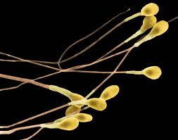
Sperm cell
|
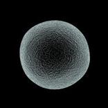
Egg cell
|
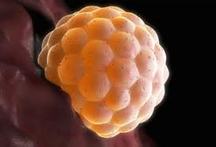
Fertilized Egg
|
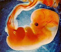
Fetus
|
Credits to the owner: https://8salembodysystems.wikispaces.com/Reproductive+System
