
Body Tissues
Tissue is a group of cells that have similar structure and that function together as a unit. A nonliving material, called the intercellular matrix, fills the spaces between the cells. This may be abundant in some tissues and minimal in others. The intercellular matrix may contain special substances such as salts and fibers that are unique to a specific tissue and gives that tissue distinctive characteristics. There are four main tissue types in the body: epithelial, connective, muscle, and nervous. Each is designed for specific functions. Use the hyperlinks below to branch into a tissue type and learn more about the topic.
Tissue is a group of cells that have similar structure and that function together as a unit. A nonliving material, called the intercellular matrix, fills the spaces between the cells. This may be abundant in some tissues and minimal in others. The intercellular matrix may contain special substances such as salts and fibers that are unique to a specific tissue and gives that tissue distinctive characteristics. There are four main tissue types in the body: epithelial, connective, muscle, and nervous. Each is designed for specific functions. Use the hyperlinks below to branch into a tissue type and learn more about the topic.
- Epithelial Tissue
- Connective Tissue
- Muscle Tissue
- Nervous Tissue
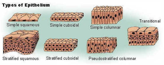
Epithelial Tissues
Epithelial tissues are widespread throughout the body. They form the covering of all body surfaces, line body cavities and hollow organs, and are the major tissue in glands. They perform a variety of functions that include protection, secretion, absorption, excretion, filtration, diffusion, and sensory reception.
The cells in epithelial tissue are tightly packed together with very little intercellular matrix. Because the tissues form coverings and linings, the cells have one free surface that is not in contact with other cells. Opposite the free surface, the cells are attached to underlying connective tissue by a non-cellular basement membrane. This membrane is a mixture of carbohydrates and proteins secreted by the epithelial and connective tissue cells.
Epithelial cells may be squamous, cuboidal, or columnar in shape and may be arranged in single or multiple layers.
Simple cuboidal epithelium is found in glandular tissue and in the kidney tubules. Simple columnar epithelium lines the stomach and intestines. Pseudostratified columnar epithelium lines portions of the respiratory tract and some of the tubes of the male reproductive tract. Transitional epithelium can be distended or stretched. Glandular epithelium is specialized to produce and secrete substances.
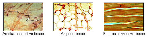
Connective Tissues
Connective tissues bind structures together, form a framework and support for organs and the body as a whole, store fat, transport substances, protect against disease, and help repair tissue damage. They occur throughout the body. Connective tissues are characterized by an abundance of intercellular matrix with relatively few cells. Connective tissue cells are able to reproduce but not as rapidly as epithelial cells. Most connective tissues have a good blood supply but some do not.
Numerous cell types are found in connective tissue. Three of the most common are the fibroblast,macrophage, and mast cell. The types of connective tissue include loose connective tissue, adipose tissue, dense fibrous connective tissue, elastic connective tissue, cartilage, osseous tissue (bone), and blood.
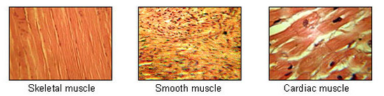
Muscle Tissue
Muscle tissue is composed of cells that have the special ability to shorten or contract in order to produce movement of the body parts. The tissue is highly cellular and is well supplied with blood vessels. The cells are long and slender so they are sometimes called muscle fibers, and these are usually arranged in bundles or layers that are surrounded by connective tissue. Actin and myosin are contractile proteins in muscle tissue.
Muscle tissue can be categorized into skeletal muscle tissue, smooth muscle tissue, and cardiac muscle tissue.
Skeletal muscle fibers are cylindrical, multinucleated, striated, and under voluntary control. Smooth muscle cells are spindle shaped, have a single, centrally located nucleus, and lack striations. They are called involuntary muscles. Cardiac muscle has branching fibers, one nucleus per cell, striations, and intercalated disks. Its contraction is not under voluntary control.
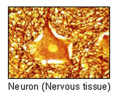
NervousTissue
Nervous tissue is found in the brain, spinal cord, and nerves. It is responsible for coordinating and controlling many body activities. It stimulates muscle contraction, creates an awareness of the environment, and plays a major role in emotions, memory, and reasoning. To do all these things, cells in nervous tissue need to be able to communicate with each other by way of electrical nerve impulses.
The cells in nervous tissue that generate and conduct impulses are called neurons or nerve cells. These cells have three principal parts: the dendrites, the cell body, and one axon. The main part of the cell, the part that carries on the general functions, is the cell body. Dendrites are extensions, or processes, of the cytoplasm that carry impulses to the cell body. An extension or process called an axon carries impulses away from the cell body.
Nervous tissue also includes cells that do not transmit impulses, but instead support the activities of the neurons. These are the glial cells (neuroglial cells), together termed the neuroglia. Supporting, or glia, cells bind neurons together and insulate the neurons. Some are phagocytic and protect against bacterial invasion, while others provide nutrients by binding blood vessels to the neurons.
Body Membranes
Body membranes are thin sheets of tissue that cover the body, line body cavities, and cover organs within the cavities in hollow organs. They can be categorized into epithelial and connective tissue membrane.Epithelial MembranesEpithelial membranes consist of epithelial tissue and the connective tissue to which it is attached. The two main types of epithelial membranes are the mucous membranes and serous membranes
Mucous Membranes. Mucous membranes are epithelial membranes that consist of epithelial tissue that is attached to an underlying loose connective tissue. These membranes, sometimes called mucosae, line the body cavities that open to the outside. The entire digestive tract is lined with mucous membranes. Other examples include the respiratory, excretory, and reproductive tracts.
Serous Membranes Serous membranes line body cavities that do not open directly to the outside, and they cover the organs located in those cavities. Serous membranes are covered by a thin layer of serous fluid that is secreted by the epithelium. Serous fluid lubricates the membrane and reduces friction and abrasion when organs in the thoracic or abdominopelvic cavity move against each other or the cavity wall. Serous membranes have special names given according to their location. For example, the serous membrane that lines the thoracic cavity and covers the lungs is called pleura.
Connective Tissue MembranesConnective tissue membranes contain only connective tissue. Synovial membranes and meninges belong to this category.
Synovial Membranes. Synovial membranes are connective tissue membranes that line the cavities of the freely movable joints such as the shoulder, elbow, and knee. Like serous membranes, they line cavities that do not open to the outside. Unlike serous membranes, they do not have a layer of epithelium. Synovial membranes secrete synovial fluid into the joint cavity, and this lubricates the cartilage on the ends of the bones so that they can move freely and without friction.
Meninges. The connective tissue covering on the brain and spinal cord, within the dorsal cavity, are called meninges. They provide protection for these vital structures.
Credits: http://free-ed.net/free-ed/Resources/Sci/Biol/AnatomyPhysiol/Human01.asp?iNum=0
